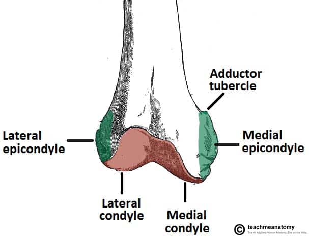
Called also lateral epicondyle b. The fibular head forms a fibrous connective tissue.

Leg Knee Anatomy
At its upper part is the.
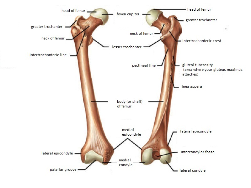
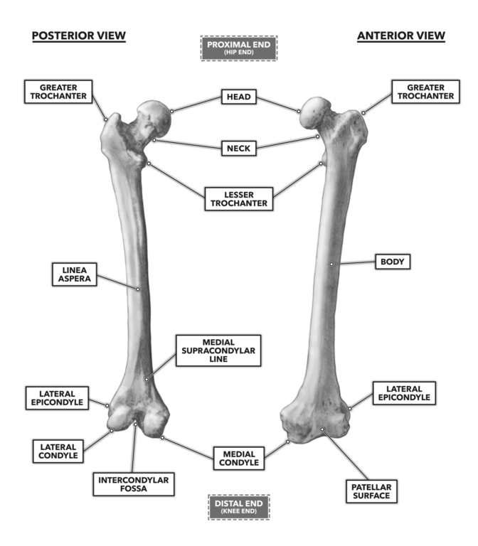
. Lateral and medial condyles. Posteriorly these condyles are separated by the deep intercondylar fossa. On the posterior surface of the condyle the linea aspera a ridge with two lips.
The medial condyle is one of the two projections on the lower extremity of femur the other being the lateral condyle. Bucket handle semilunar cartilage - see. NOTES NOTES BONES JOINTS.
The medial epicondyle is a large convex eminence to which the medial collateral ligament of the knee-joint is attached. Breast bone - see Fracture sternum. Distally on the femur are the lateral and medial condyles which articulate with the tibia below.
Following insertion of orthopedic implant joint prosthesis or bone plate - see Fracture following insertion of orthopedic implant joint prosthesis or bone plate. There are two femoral condyles medial and lateral that articulate with the medial and lateral menisci two c-shaped fibrocartilage structures that cover the tibial condyle and alleviate the incongruity between the two surfaces. A larger and more prominent one on the inner aspect of the distal part of the humerus or proximal to the medial condyle of.
Metacarpals phalanges hands fingers. The femoral and tibial condyles barely directly articulate in the normal stifle because of the menisci. The lower extremity of femur or distal.
Epicondyles Each condyle is surmounted by an elevation the epicondyle. Pathological cause unknown - see Fracture pathological. Anteriorly on the distal femur is the smooth patellar surface which forms a joint with the patella or kneecap.
80 bones 22 in skull 33 vertebrae 24 ribs 1 sternum Long bones Length width Humerus radius ulna in arms. Of the medial wall of the fossa and the anterior cruciate ligament to an impression on the upper and back part of its lateral wall. In due to - see Fracture pathological due to neoplastic disease.
The medial condyle is larger than the lateral outer condyle due to more weight bearing caused by the centre of mass being medial to the knee.
The Femur Proximal Distal Shaft Teachmeanatomy
Femur Anatomy And Attachments Bone And Spine

Epicondyles And Condyles Are Sites Of Muscle And Ligament Attachment Epicondyles Are Raised Above A Condyle Muscle
Crossfit Bones Of The Knee

Bones The Knee Doc
Orif Lag Screw For Lateral Medial Femoral Epicondyle Fracture

Femur An Overview Sciencedirect Topics
What Is The Difference Between Condyle And Epicondyle Quora
0 comments
Post a Comment